Phage Display Selection, In Vitro Characterization, and Correlative PET Imaging of a Novel HER3 Peptide
2018 April
ABSTRACT
Purpose
HER3 (ERBB3) is a receptor tyrosine kinase that is implicated in treatment resistance across multiple cancers, including those of the breast, lung, and prostate. Overexpression of HER3 following targeted therapy can occur rapidly and heterogeneously both within a single lesion and across sites of metastasis, making protein quantification by biopsy highly challenging. A global, non-invasive methodology such as positron emission tomography (PET) imaging can permit serial quantification of HER3, providing a useful approach to monitor HER3 expression across the entire tumor burden both prior to and following treatment. PET imaging of HER3 expression may permit a more personalized approach to targeted therapy by allowing for detection of HER3-mediated resistance, in addition to informing clinical trial patient selection for novel therapies targeting HER3.
Procedures
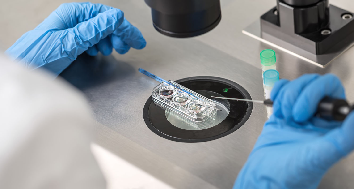
Phage display selection targeting the HER3 extracellular domain was performed in order to develop a peptide with optimal blood clearance and highly accurate HER3 quantification.
Results
The selection converged to a consensus peptide sequence that was subsequently found to bind HER3 with an affinity of 270 ± 151 nM. The peptide, termed HER3P1, was bound with high selectivity to HER3 over other similar receptor tyrosine kinases such as EGFR and HER2. Furthermore, HER3P1 was able to distinguish between high and low HER3-expressing cells in vitro. The peptide was radiolabeled with Ga-68 and demonstrated to specifically bind HER3 by in vivo PET imaging. Uptake of [68Ga]HER3P1 was highly specific for HER3-positive tumors, with tumor-to-background ratios ranging from 1.59–3.32, compared to those of HER3-negative tumors, ranging from 0.84–0.93. The uptake of [68Ga]HER3P1 also demonstrated high (P < 0.001) correlation with protein expression as quantified by Western blot and confirmed by biodistribution.
Conclusions
HER3P1 accurately quantifies expression of HER3 by PET imaging and has potential utility as a clinical imaging agent.
Key words
HER3 PET Phage display Peptide
NOTES
Acknowledgements
We would like to thank Emily Bloch, Sarah Nesti, and Catharina Dekker for technical assistance and manuscript preparation. Funding provided by a Department of Defense Prostate Cancer Research Postdoctoral Training Award W81XWH-16-1-0447 and a Department of Defense Prostate Cancer Synergistic Idea Development Award W81XWH-14-1-0406.
Compliance with Ethical Standards
Conflict of Interest
The authors declare that they have no conflict of interest.
REFERENCES
-
Herbst RS, Prager D, Hermann R et al (2005) TRIBUTE: a phase III trial of erlotinib hydrochloride (OSI-774) combined with carboplatin and paclitaxel chemotherapy in advanced non-small-cell lung cancer. J Clin Oncol 23:5892–5899CrossRefPubMedGoogle Scholar
-
Chapman PB, Hauschild A, Robert C et al (2011) Improved survival with vemurafenib in melanoma with BRAF V600E mutation. N Engl J Med 364:2507–2516CrossRefPubMedPubMedCentralGoogle Scholar
-
Engelman JA, Zejnullahu K, Mitsudomi T et al (2007) MET amplification leads to gefitinib resistance in lung cancer by activating ERBB3 signaling. Science 316:1039–1043CrossRefPubMedGoogle Scholar
-
Sergina NV, Rausch M, Wang D et al (2007) Escape from HER-family tyrosine kinase inhibitor therapy by the kinase-inactive HER3. Nature 445:437–441CrossRefPubMedPubMedCentralGoogle Scholar
-
Chandarlapaty S (2012) Negative feedback and adaptive resistance to the targeted therapy of cancer. Cancer Discov 2:311–319CrossRefPubMedPubMedCentralGoogle Scholar
-
Schlessinger J (2000) Cell signaling by receptor tyrosine kinases. Cell 103:211–225CrossRefPubMedGoogle Scholar
-
Yarden Y (2001) Untangling the ErbB signalling network. Nat Rev Mol Cell Biol 2:127CrossRefPubMedGoogle Scholar
-
Ferguson KM, Darling PJ, Mohan MJ et al (2000) Extracellular domains drive homo- but not hetero-dimerization of erbB receptors. EMBO J 19:4632–4643CrossRefPubMedPubMedCentralGoogle Scholar
-
Slamon DJ, Leyland-Jones B, Shak S et al (2001) Use of chemotherapy plus a monoclonal antibody against HER2 for metastatic breast cancer that overexpresses HER2. N Engl J Med 344:783–792CrossRefPubMedGoogle Scholar
-
Franklin MC, Carey KD, Vajdos FF et al (2004) Insights into ErbB signaling from the structure of the ErbB2-pertuzumab complex. Cancer Cell 5:317–328CrossRefPubMedGoogle Scholar
-
Shi F, Telesco SE, Liu Y et al (2010) ErbB3/HER3 intracellular domain is competent to bind ATP and catalyze autophosphorylation. Proc Natl Acad Sci U S A 107:7692–7697CrossRefPubMedPubMedCentralGoogle Scholar
-
Chakrabarty A, Sánchez V, Kuba MG et al (2012) Feedback upregulation of HER3 (ErbB3) expression and activity attenuates antitumor effect of PI3K inhibitors. Proc Natl Acad Sci U S A 109:2718–2723CrossRefPubMedGoogle Scholar
-
Soler M, Mancini F, Meca-Cortés Ó et al (2009) HER3 is required for the maintenance of neuregulin-dependent and -independent attributes of malignant progression in prostate cancer cells. Int J Cancer 125:2565–2575CrossRefPubMedGoogle Scholar
-
Chen L, Siddiqui S, Bose S et al (2010) Nrdp1-mediated regulation of ErbB3 expression by the androgen receptor in androgen-dependent but not castrate-resistant prostate cancer cells. Cancer Res 70:5994–6003CrossRefPubMedPubMedCentralGoogle Scholar
-
Lidke DS, Nagy P, Heintzmann R et al (2004) Quantum dot ligands provide new insights into erbB/HER receptor-mediated signal transduction. Nat Biotech 22:198–203CrossRefGoogle Scholar
-
Heidari P, Wehrenberg-Klee E, Habibollahi P et al (2013) Free somatostatin receptor fraction predicts the antiproliferative effect of octreotide in a neuroendocrine tumor model: implications for dose optimization. Cancer Res 73:6865–6873CrossRefPubMedGoogle Scholar
-
Fischman AJ, Babich JW, Strauss HW (1993) A ticket to ride: peptide radiopharmaceuticals. J Nucl Med 34:2253–2263PubMedGoogle Scholar
-
Hofmann M, Maecke H, Börner A et al (2001) Biokinetics and imaging with the somatostatin receptor PET radioligand 68Ga-DOTATOC: preliminary data. Eur J Nucl Med Mol Imaging 28:1751–1757CrossRefGoogle Scholar
-
Poeppel TD, Binse I, Petersenn S et al (2011) 68Ga-DOTATOC versus 68Ga-DOTATATE PET/CT in functional imaging of neuroendocrine tumors. J Nucl Med 52:1864–1870CrossRefPubMedGoogle Scholar
-
Smith GP (1985) Filamentous fusion phage: novel expression vectors that display cloned antigens on the virion surface. Science 228:1315–1317CrossRefPubMedGoogle Scholar
-
Karasseva NG, Glinsky VV, Chen NX et al (2002) Identification and characterization of peptides that bind human ErbB-2 selected from a bacteriophage display library. J Prot Chem 21:287–296CrossRefGoogle Scholar
-
Larimer BM, Thomas WD, Smith GP, Deutscher SL (2014) Affinity maturation of an ERBB2-targeted SPECT imaging peptide by in vivo phage display. Mol Imaging Biol 16:449–458CrossRefPubMedGoogle Scholar
-
Ruoslahti E (1996) RGD and other recognition sequences for integrins. Annu Rev Cell Dev Biol 12:697–715CrossRefPubMedGoogle Scholar
-
Larimer BM, Deutscher SL (2014) Development of a peptide by phage display for SPECT imaging of resistance-susceptible breast cancer. Am J Nuc Med Mol Imaging 4:435Google Scholar
-
Wakui H, Yamamoto N, Nakamichi S et al (2014) Phase 1 and dose-finding study of patritumab (U3-1287), a human monoclonal antibody targeting HER3, in Japanese patients with advanced solid tumors. Cancer Chemother Pharmacol 73:511–516CrossRefPubMedPubMedCentralGoogle Scholar
-
Deutscher SL (2010) Phage display in molecular imaging and diagnosis of cancer. Chem Rev 110:3196–3211CrossRefPubMedPubMedCentralGoogle Scholar
-
Thomas WD, Golomb M, Smith GP (2010) Corruption of phage display libraries by target-unrelated clones: diagnosis and countermeasures. Anal Biochem 407:237–240CrossRefPubMedPubMedCentralGoogle Scholar
-
Lockhart AC, Liu Y, Dehdashti F et al (2016) Phase 1 evaluation of [64Cu]DOTA-patritumab to assess dosimetry, apparent receptor occupancy, and safety in subjects with advanced solid tumors. Mol Imaging Biol 18:446–453CrossRefPubMedPubMedCentralGoogle Scholar
-
Wehrenberg-Klee E, Turker NS, Heidari P et al (2016) Differential receptor tyrosine kinase PET imaging for therapeutic guidance. J Nucl Med 57:1413–1419CrossRefPubMedPubMedCentralGoogle Scholar
-
Da Pieve C, Allott L, Martins CD et al (2016) Efficient [18F]AlF radiolabeling of ZHER3:8698 affibody molecule for imaging of HER3 positive tumors. Bioconjug Chem 27:1839–1849CrossRefPubMedGoogle Scholar
-
Wang M, Gao M, Zheng Q-H (2014) The first radiosynthesis of [11 C] AZD8931 as a new potential PET agent for imaging of EGFR, HER2 and HER3 signaling. Bioorg Med Chem Lett 24:4455–4459CrossRefPubMedGoogle Scholar